Applied Physics, Engineering, and Biology
As an undergraduate and masters student in the Quantum Optics and Laser Science Group at Imperial College, I studied the interactions of lasers with matter under the direction of Profs. Jon Marangos and Roland Smith. My interests in developing new technologies led me to deltaDOT, a DNA-sequencing startup that originated in the Imperial College Dept. of High Energy Physics. My experience at deltaDOT was formative for me in viewing biology as the technological future.
As a graduate student at Cornell University, I was drawn by the cutting-edge visions for biological science that would be made possible at the Energy Recovery Linac (ERL), a new type of particle accelerator that was first conceived at Cornell. Joining a team of professors, postdocs, and staff scientists on the Cornell ERL Project, I worked on the design of higher order mode absorbers under Profs. Sol Gruner and Hasan Padamsee. Following this work, I became increasingly interested in the biological studies that are enabled by synchrotron X-rays and pursued my dissertation research in protein biophysics with Prof. Sol Gruner. Using X-ray crystallography, high-pressure engineering, and optical spectroscopy, I correlated sub-angstrom changes in protein structure to large functional changes. During this time, I was also one of the students that were involved in building G-line, the newest addition to the Cornell High Energy Synchrotron Source (CHESS).
My graduate research on protein structure-function steered me towards protein design and biological engineering in my postdoctoral work. As a postdoctoral fellow in Prof. Pamela Silver's group, I am currently using the methods of synthetic biology to tackle problems in bioenergy.
A. Synthetic approaches to biological energy production
The world faces a crisis of energy, sustainability and climate in the coming century. In the next twenty-five years, world energy use is projected to increase from 17 TW to 26 TW [1]. If this energy is supplied by fossil sources, it will pollute the atmosphere with carbon dioxide with consequences for centuries to come. Even if this carbon dioxide is sequestered, we are still faced with a tightening in the production of coal, the most abundant fossil fuel, before 2050. Nature has thus far solved the problems of non-polluting energy capture and storage at the scale needed by civilization, channeling ~100 TW by photosynthesis into biomass [2]. However, the average photosynthetic conversion of sunlight to biomass rarely exceeds 1% in efficiency [3]. Can we better channel solar energy into non-polluting fuel production using the methods of synthetic biology? One tantalizing approach is to artificially link photosynthetic solar energy capture to the biological production of hydrogen gas, a zero-emission fuel. The greatest hurdle to this approach has been that hydrogen-producing enzymes (hydrogenases) with the highest activity use iron cofactors that are inactivated by the oxygen produced by photosynthesis. Oxygen-tolerant properties can be engineered into hydrogenases with a high-throughput approach if these enzymes are evolved in microbial hosts that are undergoing a genetic selection pressure. A newly developed method links hydrogenase activity to essential sulfur metabolism in E. coli, such that only hosts that develop oxygen-tolerant mutations to the hydrogenase gene can survive [4].
[1] Conti and Holtberg. International Energy Outlook 2011.
[2] Whittaker and Likens. 1975. "The Biosphere and Man". In "Primary Productivity of the Biosphere". Berlin: Springer-Verlag. pp. 305–328.
[3] Blankenship, Tiede, Barber, Brudvig, Fleming, et al. Science (2011) 332: 805–809.
[4] Barstow, Agapakis, Boyle, Grandl, Silver and Wintermute. J. of Biol. Eng. (2011), 5:7.
B. Protein structure-function at the sub-angstrom scale
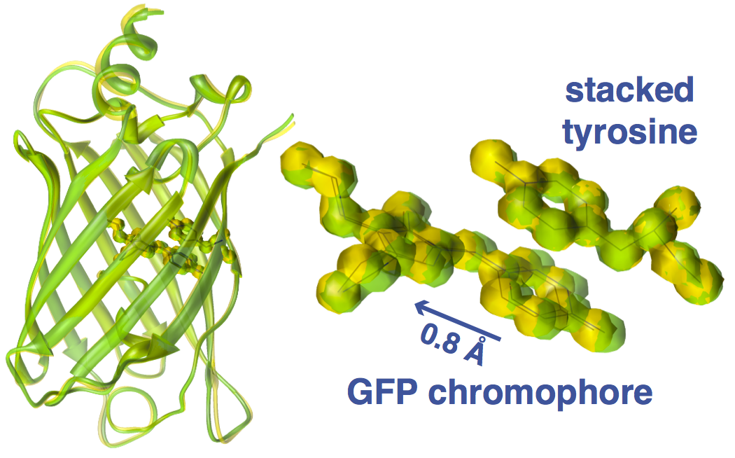
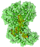
The structure of a protein is crucial for its function, but at what length scales does atomic positioning matter for protein function? As proteins have dimensions of tens to hundreds of angstroms, sub-angstrom changes to atomic positions may at first seem insignificant. However, if one considers two objects that are separated by 3.5 Å interacting through a Lennard-Jones potential and moves them closer together by just 0.1 Å, the potential energy between these objects changes by 19%! Thus, changes as small as 0.1 Å can make a large difference in the energy of an electronic transition, and thus a notable change in protein function. Given this result, what is the detection limit of small structural motions in proteins? While changes on the order of 0.1 Å are generally difficult to resolve in crystallography, they can be detected if multiple snapshots can be obtained depicting a progressive change and if the structural changes are larger than experimental standard deviations or the estimated coordinate error [1,2]. Sensitivity to such sub-angstrom structural perturbations is observed in citrine, a yellow variant of the Green Fluorescent Protein (GFP) [1]. Citrine differs from GFP in that a tyrosine is stacked 3.5 Å above the GFP chromophore, which perturbs the excited state of the chromophore and shifts the fluorescence from green to yellow. Very small structural changes can be introduced by pressurizing crystals and then locked into place by freezing [3]. The resultant series of 26 high-resolution crystal structures depict the GFP chromophore gradually sliding away from the stacked tyrosine over ~0.8 Å (with standard deviations of replicate data less than or equal to the estimated coordinate error of 0.2 Å) [1,2]. Amazingly, this minuscule change in ring stacking is sufficient to cause an enormous shift in fluorescence from yellow to green, reverting citrine to a GFP-like state [1,2]. Why does the GFP chromophore slide away from the stacked tyrosine when citrine is pressurized? The reason is that proteins are not uniformly compressible - some regions are squishier than others. Thus, an overall compression can lead to some residues moving apart from each other. In the case of citrine, the GFP chromophore and stacked tyrosine are parts of two separate clusters of residues that move relative to each other under hydrostatic pressure, causing the rings to slide apart [4].
[1] Barstow, Ando, Kim and Gruner. PNAS (2008) 105: 13362-13366.
[2] Ando and Barstow. Enc. Anal. Chem. (2012) doi: 10.1002/9780470027318.a9246.
[3] Kim, Kapfer, and Gruner. Acta Crystallogr D (2005) 61:881– 890.
[4] Barstow, Ando, Kim and Gruner. Biophysical Journal (2009), 97: 1719–1727.
[1] Barstow, Ando, Kim and Gruner. PNAS (2008) 105: 13362-13366.
[2] Ando and Barstow. Enc. Anal. Chem. (2012) doi: 10.1002/9780470027318.a9246.
[3] Kim, Kapfer, and Gruner. Acta Crystallogr D (2005) 61:881– 890.
[4] Barstow, Ando, Kim and Gruner. Biophysical Journal (2009), 97: 1719–1727.
C. A new type of synchrotron based on energy recovery

Synchrotrons support an enormous amount of science in many areas, from biology to materials science to geology. These machines make use of the fact that charged particles accelerating at high energies radiate light, including X-rays, at many useful wavelengths. By radiating light, however, the charged particles also lose energy. Traditionally, synchrotrons have relied on storage rings to restore energy into the orbiting particles. In contrast, the Energy Recovery Linac (ERL) is a radically different type of particle accelerator that was first envisioned by Maury Tigner at Cornell [1]. Instead of recycling particles, the ERL will recycle energy by dumping particles after every orbit and recovering their kinetic energy for accelerating the next bunch of charged particles. By doing so, charged particles maintain their small bunch structure, which in turn allows for X-ray beams with unprecedented characteristics such as a ultrashort pulses, high brilliance, nano beam sizes, and very high repetition rates [2]. The particles in the ERL will be accelerated by an electromagnetic field generated by superconducting radio-frequency (RF) cavities [3]. One of the most important aspects to maintaining the stability of the particles bunches is to make sure that higher order modes are dampened in the resonant RF cavities [4,5]. Importantly, the ERL maintains the ring structure of traditional synchrotrons and therefore, will service many users at once. With the ERL, state-of-the-art science can be imagined, such as determining protein structures without crystals with fast data acquisition and exploring protein structural dynamics at short timescales.
[1] Tigner. Nuovo Cimento (1965) 7: 1228–1231.
[2] Bilderback, Brock, Dale, Finkelstein, Pfeifer, and Gruner. New J. Physics (2010) 12, 035011.
[3] Hoffstaetter, Barstow, et al. Proc. of Particle Accelerator Conference (2003), 1: 192-194.
[4] Liepe, Barstow, Padamsee. Proc. of Particle Accelerator Conference (2003), 2: 1320-1322.
[5] Barstow, Liepe, Padamsee. Proc. of Workshop on Superconducting Radio Frequency (2003).
